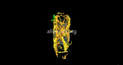
This 3-D model of how cells divide may lead to breakthroughs in cancer research

Cell division is a process that scientists have been fascinated with since we first learned about cells. Through decades of study, scientists have come up with a basic narrative on how cells divide: Each phase of division, broadly called "mitosis," has been catalogued and analyzed up close. Now, the Allen Institute for Cell Science has come up with a better way to take a good look at the way all organisms form: a 3-D model that visualizes, in color-coded detail, the way a healthy human cell divides.
Announced in a press release on Wednesday, the Allen Institute's model of the Integrated Mitotic Stem Cell will enable "a deeper understanding" of the process of mitosis in human cells. In addition to helping us with "basic biology research," it will also be instrumental in cancer-related research.
Cancer is caused by the improper division and replication of cells — in the search for treatments and cures, scientists are often looking at why the specific cells that make up a cancerous growth are behaving that way. So having a full model of how a normal, healthy cell divides provides "a much-needed baseline" for comparison to cancerous cells, said Rick Horwitz, the executive director of the Allen Institute's Cell Science division.
Subscribe to The Week
Escape your echo chamber. Get the facts behind the news, plus analysis from multiple perspectives.

Sign up for The Week's Free Newsletters
From our morning news briefing to a weekly Good News Newsletter, get the best of The Week delivered directly to your inbox.
From our morning news briefing to a weekly Good News Newsletter, get the best of The Week delivered directly to your inbox.
Further studies into the mitosis process will be able to use the Allen Insitute's tool "to connect the dots between different parts of the cell," instead of studying just the chromosomes in isolation, said Tom Misteli, the director of the National Cancer Institute's Center for Cancer Research.
Take a look at the Integrated Mitotic Stem Cell here, or watch the Allen Institute's video about it below. Shivani Ishwar
Create an account with the same email registered to your subscription to unlock access.
Sign up for Today's Best Articles in your inbox
A free daily email with the biggest news stories of the day – and the best features from TheWeek.com
Shivani is the editorial assistant at TheWeek.com and has previously written for StreetEasy and Mic.com. A graduate of the physics and journalism departments at NYU, Shivani currently lives in Brooklyn and spends free time cooking, watching TV, and taking too many selfies.
-
 Today's political cartoons - April 13, 2024
Cartoons Saturday's cartoons - moderate MAGA, automotive politics, and more
By The Week US Published
-
 5 Grand Canyon-size cartoons on the Arizona abortion ruling
Cartoons Artists take on a chasm in reproductive freedom, the dangers of an abortion ban, and more
By The Week US Published
-
 Crossword: April 13, 2024
The Week's daily crossword
By The Week Staff Published
-
 Puffed rice and yoga: inside the collapsed tunnel where Indian workers await rescue
Speed Read Workers trapped in collapsed tunnel are suffering from dysentery and anxiety over their rescue
By Sorcha Bradley, The Week UK Published
-
 More than 2,000 dead following massive earthquake in Morocco
Speed Read
By Justin Klawans Published
-
 Mexico's next president will almost certainly be its 1st female president
Speed Read
By Peter Weber Published
-
 North Korea's Kim to visit Putin in eastern Russia to discuss arms sales for Ukraine war, U.S. says
Speed Read
By Peter Weber Published
-
 Gabon's military leader sworn in following coup in latest African uprising
Speed Read
By Justin Klawans Published
-
 Nobody seems surprised Wagner's Prigozhin died under suspicious circumstances
Speed Read
By Peter Weber Published
-
 Western mountain climbers allegedly left Pakistani porter to die on K2
Speed Read
By Justin Klawans Published
-
 'Circular saw blades' divide controversial Rio Grande buoys installed by Texas governor
Speed Read
By Peter Weber Published